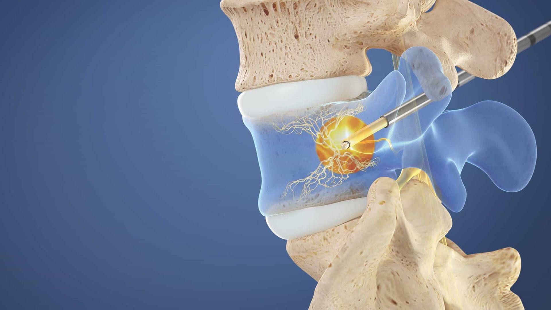
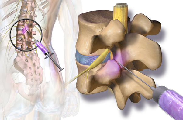
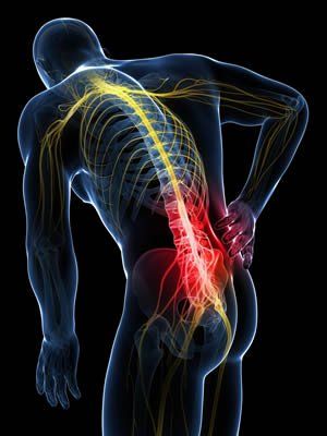
Blog Layout

Services

Acupuncture For Chronic Pain

I recently reviewed a report on acupuncture for chronic pain. It reviewed 13 clinical randomized trials that occurred up until December 2015 and included over 20,000 patients. It compared the use of acupuncture to no acupuncture in patients or sham acupuncture, which was fake acupuncture and it found a significant difference in both pain relief and improved function in the patients using real acupuncture. It also found that the results can persist over many months.


Our clinic does feel that acupuncture can be a significant beneficial modality for treatment of many types of pain. Patients can sometimes lower their pain medication with the use of acupuncture. Some insurances do cover it. If you would like to discuss this with your provider let us know.

Conditions & Procedures

Spine and Nerve RSS Feed

04 Nov, 2020
A sacroiliac joint (SI) injection is an image guided, minimally invasive method of targeted drug therapy which is performed to relieve low back/buttock pain. Steroid medication can reduce the swelling and inflammation caused by conditions such as arthritis or joint dysfunction. This minimizes side effects when compared to oral medications and increases efficacy. How is a sacroiliac joint injection performed? Patient Positioning The patient lies face down. Tissue Anesthetized A local anesthetic (typically lidocaine) is used to numb the skin and the tissues along the anticipated path of the needle. Fluoroscopic Guidance Using a fluoroscope for guidance (live x-ray guidance), the physician directs the needle toward the sacroiliac joint. A contrast solution is injected to assess for ideal location and safety. The physician uses the fluoroscope to confirm the correct location of the needle tip. Steroids Injected A steroid-anesthetics mix is injected into the sacroiliac joint, bathing the painful tissues with a potent anti-inflammatory medication. End of Procedure The needle is removed and a small bandage is applied to cover the tiny needle surface wound. How long does it take for the sacroiliac joint injection to work? Most people feel some benefit from the injection between 3-7 days however it can take up to two weeks before we see the full results. Who performs a sacroiliac joint injection? Sacroiliac joint injections are performed by one of the board certified interventionalists at Spine & Nerve Diagnostic Center (SNDC). Our interventionalists have performed thousands of these procedures. Where are sacroiliac joint injections performed? Sacroiliac joint injections are typically performed at our state of the art SNDC procedure suites.
04 Nov, 2020
Overview: Whiplash happens when your neck jerks back and forth quickly and violently, causing your spine to bend past its normal range of motion. This can injure the vertebrae of your cervical spine and/or the ligaments and muscles in your neck. Causes: Whiplash is commonly caused by rear-end collision motor vehicle accidents or by contact sports such as football. Symptoms: You may feel pain and stiffness in your neck, shoulders, back, and arms at the time of your injury, or it may begin days later. Treatment Options: Treatment options may include rest, physical therapy, medications and injections.
04 Nov, 2020
Overview The rotator cuff of the shoulder is composed of four different muscles that support the shoulder joint. This group of muscles keeps the head of the humerus (upper arm bone) secured within the shallow socket of the shoulder joint. When the rotator cuff tendons are injured they become irritated or damaged, resulting in pain, weakness and reduced range of motion. Rotator cuff injuries are common and increase with age, in people who have jobs with overhead motions such as painters and carpenters. Many people can manage their symptoms and return to activities with physical therapy to improve strength and flexibility; however some patients also require surgical interventions to help correct the injury. Symptoms Rotator cuff injuries can manifest as pain, weakness, and/or range of motion. Pain in rotator cuff injuries can be described as a deep ache in the shoulder that can also disturb sleep. Rotator cuff injuries will make it difficult for the person to perform any over head motions such as brushing your hair, or reaching behind your back. Patients may also experience weakness when performing specific movements. Diagnosis Diagnosis of rotator cuff injuries is primarily through physical exam with your medical provider. X-rays, ultrasound, and MRI are also utilized to help diagnose the extent of rotator cuff injuries. Treatment Conservative treatments for rotator cuff injuries include rest, ice, and physical therapy. If these do not provide relief then cortisone or steroid injection can be helpful in reducing inflammation in the shoulder joint. As a last resort corrective surgery may be needed to repair the rotator cuff tendons, this is typically done arthroscopically.
04 Nov, 2020
Overview Knee osteoarthritis is a degenerative disease involving the cartilage in the knee joint. Osteoarthritis occurs when the protective cartilage surrounding the end of your bone begins to wear down. It can most commonly affect the knees, hips, hands, and spine. Symptoms can usually be managed, but the damage to the joints can never be reversed. Risk factors for developing osteoarthritis include age, gender, obesity, previous joint injuries, repetitive stress on a joint, genetics, bone deformities, and metabolic disorders. Symptoms Symptoms of osteoarthritis develop slowly over time and can vary from person to person. Most commonly, patients will experience joint pain, stiffness (especially in the morning), tenderness around the joint, loss of flexibility or range of motion, grinding sensation, and swelling. Diagnosis Diagnosis of osteoarthritis of a joint can be made by taking an x-ray of the joint to evaluate the bones in the knee joint. An MRI may also be useful in diagnosing osteoarthritis to help evaluate the soft tissue around the joint. Treatment Treatment of osteoarthritis first begins with lifestyle changes including consistent exercise and proper weight management. Physical therapy, and pool therapy have also been shown to be effective in helping treat osteoarthritis pain. Additionally, alternative movement therapies such as tai-chi and yoga have been found to help reduce pain caused by osteoarthritis. Traditional medication options include acetaminophen (Tylenol), and non-steroidal anti-inflammatory drugs (NSAIDs) such as ibuprofen, or Aleve. Supplements such as glucosamine/chondroitin, capsaicin, and omega-3 fatty acids, while controversial in efficacy, have anecdotally reduced patient’s osteoarthritis pain. Devices such as knee braces and a TENS unit can also provide relief. Typically, if lifestyle changes, therapies, and medications fail to relieve pain, then cortisone or steroid injections can be performed to help reduce inflammation in the knee joint. Other available injections include hyaluronic acid, prolo injection therapy, and platelet-rich-plasma injections. Finally, if all conservative measures fail, the patient can undergo surgery to change the structure of the knee. Two types of surgeries are available: partial knee replacement and total knee replacement.
04 Nov, 2020
This is a disease of the spine indicating degenerative changes, sometimes referred to as arthritis of the spine. It can happen at any level (cervical, thoracic or lumbar) and results in different types of pain including mechanical and radicular pain. CAUSES Primarily this is caused by aging and normal wear and tear. With time, the components of the spine, including the vertebrae, the discs between the vertebrae, and the joints of the spine rub against one another, causing pain. SYMPTOMS Cervical Spine: neck, shoulder and arm pain. Radicular symptoms may occur including numbness, weakness in the arms or legs. Thoracic Spine: upper chest and abdomen pain, and may cause numbness, tingling and weakness in the legs Lumbar Spine: lower back, buttocks, and leg pain; and may cause numbness, tingling and weakness in the lower leg. This is often times worsened with sitting, twisting, lifting and bending. TREATMENT This varies based on severity of degeneration, and symptoms patients describe. It includes anti- inflammatory medications, physical therapy, water therapy, back braces, spinal injections and in severe cases, surgery.
04 Nov, 2020
Facet joints are found on the back of the entire spine (cervical, thoracic and lumbar). Each vertebrae rests on top of one another at these facet joints. These joints may become inflamed or irritated, causing pain. A steroid injection into the joint may be used to help alleviate pain and diagnose the source of a patient’s pain. Q&A 1. How is a facet joint injection performed? a. Patient positioning: face down on a procedure table. 2. Will the procedure be painful? a. A local anesthetic is used to numb the skin. This feels similar to a bee sting and lasts for a very short time, usually less than 30 seconds. 3. How does the physician know where to inject? a. The physician uses fluoroscopic guidance (live XR imaging) directing the needle into the facet joint space. A contrast solution is injected and confirms the location of the medication to be placed. 4. What is injected? a. A mixture of anesthetic (numbing medicine) and steroid (anti-inflammatory) is injected into the joint space. 5. End of Procedure a. The needle is removed and a small bandage is placed to cover the site of the injection. 6. How long will it take to work? a. Relief usually takes 3-7 days to estimate relief. 7. Who performs a facet joint injection? a. Facet joint injections are performed by one of the board certified interventionalists at Spine and Nerve Diagnostic Center (SNDC). Our interventionalists have performed thousands of these procedures. 8. Where are facet joint injections performed? a. Facet joint injections are performed at our state of the art SNDC procedure suites.
04 Nov, 2020
Overview Hip osteoarthritis is a degenerative disease involving the cartilage in the hip joint. Osteoarthritis occurs when the protective cartilage surrounding the end of your bone begins to wear down over time. It can most commonly affect the knees, hips, hands, and spine. Symptoms can usually be managed but the damage to the joints can never be reversed. Risk factors for developing osteoarthritis include age, gender, obesity, previous joint injuries, repetitive stress on a joint, genetics, bone deformities, and metabolic disorders. Symptoms Symptoms of osteoarthritis develop slowly over time and can vary from person to person. Most commonly, patients will experience joint pain, stiffness (especially in the morning), tenderness around the joint, loss of flexibility or range of motion, grating sensation, and swelling. Diagnosis Diagnosis of osteoarthritis of a joint can be made with x-ray of the joint. MRI may also be useful in diagnosing osteoarthritis. Treatment Treatment of osteoarthritis first begins with lifestyle changes including consistent exercise and proper weight management. Physical therapy and pool therapy have also been shown to be effective in helping treat osteoarthritis pain. Additionally, alternative movement therapies such as tai-chi and yoga have been found to help reduce pain caused by osteoarthritis. Traditional medication options include acetaminophen (tylenol), and non-steroidal anti- inflammatory drugs (NSAIDs) such as ibuprofen, or aleve. Supplements such as glucosamine/chondroitin, capsaicin, and omega-3 fatty acids, while controversial in efficacy, have anecdotally reduced patient’s osteoarthritis pain. Typically, if lifestyle changes, therapies, and medications fail to relieve pain, then cortisone or steroid injections can be performed to help reduce inflammation in the hip joint. Finally, if all conservative measures fail the patient can undergo surgery to replace the hip joint. This is typically done by an orthopedic surgeon.
04 Nov, 2020
Piriformis syndrome occurs when the piriformis muscle, which is located deep in the buttock, causes pain. The piriformis muscle starts at the lower spine and attaches to the upper thigh bone. It functions to rotate the hip outward. What causes Piriformis Syndrome? Swelling, inflammation and spasm can occur in the piriformis muscle due to trauma, prolonged sitting or repetitive injury such as from sports. The pain may run down the back of the thigh. How is Piriformis Sydrome diagnosed? There is no direct, definitive test for Piriformis Syndrome. It is diagnosed by history and physical exam. Imaging may be ordered to rule out other pathologies. How is Piriformis Syndrome treated? Avoiding the activities that trigger the pain is important. Rest, ice, and heat can be helpful. Physical therapy, stretching, and exercises can also help relieve pain. Anti-inflammatories and muscle relaxers can ease pain. Injections with a steroid or botox can reduce the inflammation, pain, and spasm. 
04 Nov, 2020
What is medial epicondylitis? Medial Epicondylitis also know as Golfer’s elbow is the inflammation of the tendons that connect the muscles of the muscles of the forearm to the elbow. The pain is felt at the medial epicondyle (the bony bump on the inner side of the elbow). Causes of medial epicondylitis It is caused by repetitive motions, overuse of the arm, and stress on the fore arms which leads to small tears at the flexor tendons triggering pain, weakness, stiffness. Risk Factors: This condition is common among athletes such as golfers and people who play racquet sports. It also is common in persons involved in professions requiring repetitive motions such as carpenters, painters, and computer users. Symptoms of medial epicondylitis There is pain, tenderness, and stiffness at the medial epicondyle that can radiate to the fore arm and wrist. Numbness and tingling in the fingers Weakness of the hand and wrists Difficulty moving the left elbow Treatment Options Rest and avoid activities that require repetitive movement until the pain disappears. Acupuncture therapy, massage therapy and use of an electronic pulse massager can relieve the pain and improve circulation. Ice and a cold compress can help to reduce swelling and inflammation. Physical therapy/Occupational therapy: stretching helps stretch and strengthen the tendons. OTC pain killers and anti- inflammatories can help reduce the pain and inflammation. Topical ointments such as the lidopro ointment to reduced localized pain and inflammation. Steroid injections and PRP injections helps to reduce swelling, inflammation, and can initiate or expedite the healing process. Wrist splint/Brace: can provide additional support and pain relief to reduce muscle strain. Laser therapy that we offer in clinic.
28 Oct, 2020
A medial branch block is an image guided, minimally invasive method of targeted drug therapy which is performed for diagnostic purposes to confirm that the facet joints (joints in the back of the spine) are the source of your pain. Typically a medial branch block is performed twice (two separate occasions) to optimize our certainty of the diagnosis. If the blocks are positive then you may be a candidate for a procedure called a radiofrequency ablation which can provide long lasting (often 6+ months) of benefit. This procedure can be performed for the neck and the back. After the procedure is performed it is VERY important that you monitor how your pain compares to your typical pain so that you can tell your provider at the next visit. To help you remember you will be provided a pain diary to fill out following the procedure. It is important to note that this is a DIAGNOSTIC procedure and you should not expect long lasting benefits (generally no more than a few hours). It is also important to know that this procedure typically only addresses pain that stays in the neck or back and is not likely to improve pain that radiates down your arms or legs. How is a medial branch block injection performed? Patient Positioning The patient lies face down. Tissue Anesthetized A local anesthetic (typically lidocaine) is used to numb the skin. Fluoroscopic Guidance Using a fluoroscope for guidance (live x-ray guidance), the physician directs the needles to the areas where the medial branch runs towards the suspected problematic joints. The physician uses the fluoroscope to confirm the correct location of the needle tip. Medication Injected A local anesthetic is injected around the medial branch nerve which should prevent transmission of pain signals from the joint from reaching the brain for the duration that the anesthetic lasts. End of Procedure The needles are removed and a small bandage is applied to cover the tiny needle surface wound. How long does it take for the medial branch block to work? Typically the local anesthetic takes effect over the course of a few minutes and frequently you will be able to notice a difference in your symptoms as you get up off of the procedure table. As discussed above, this is a diagnostic procedure and is only expected to last for the duration of the local anesthetic. Who performs a medial branch block injection? Medial branch block injections are performed by one of the board certified interventionalists at Spine & Nerve Diagnostic Center (SNDC). Our interventionalists have performed thousands of these procedures. Where are branch block injections performed? Medial branch block injections are typically performed at our state of the art SNDC procedure suites. 
Show More
04 Nov, 2020
A sacroiliac joint (SI) injection is an image guided, minimally invasive method of targeted drug therapy which is performed to relieve low back/buttock pain. Steroid medication can reduce the swelling and inflammation caused by conditions such as arthritis or joint dysfunction. This minimizes side effects when compared to oral medications and increases efficacy. How is a sacroiliac joint injection performed? Patient Positioning The patient lies face down. Tissue Anesthetized A local anesthetic (typically lidocaine) is used to numb the skin and the tissues along the anticipated path of the needle. Fluoroscopic Guidance Using a fluoroscope for guidance (live x-ray guidance), the physician directs the needle toward the sacroiliac joint. A contrast solution is injected to assess for ideal location and safety. The physician uses the fluoroscope to confirm the correct location of the needle tip. Steroids Injected A steroid-anesthetics mix is injected into the sacroiliac joint, bathing the painful tissues with a potent anti-inflammatory medication. End of Procedure The needle is removed and a small bandage is applied to cover the tiny needle surface wound. How long does it take for the sacroiliac joint injection to work? Most people feel some benefit from the injection between 3-7 days however it can take up to two weeks before we see the full results. Who performs a sacroiliac joint injection? Sacroiliac joint injections are performed by one of the board certified interventionalists at Spine & Nerve Diagnostic Center (SNDC). Our interventionalists have performed thousands of these procedures. Where are sacroiliac joint injections performed? Sacroiliac joint injections are typically performed at our state of the art SNDC procedure suites.
04 Nov, 2020
Overview: Whiplash happens when your neck jerks back and forth quickly and violently, causing your spine to bend past its normal range of motion. This can injure the vertebrae of your cervical spine and/or the ligaments and muscles in your neck. Causes: Whiplash is commonly caused by rear-end collision motor vehicle accidents or by contact sports such as football. Symptoms: You may feel pain and stiffness in your neck, shoulders, back, and arms at the time of your injury, or it may begin days later. Treatment Options: Treatment options may include rest, physical therapy, medications and injections.
04 Nov, 2020
Overview The rotator cuff of the shoulder is composed of four different muscles that support the shoulder joint. This group of muscles keeps the head of the humerus (upper arm bone) secured within the shallow socket of the shoulder joint. When the rotator cuff tendons are injured they become irritated or damaged, resulting in pain, weakness and reduced range of motion. Rotator cuff injuries are common and increase with age, in people who have jobs with overhead motions such as painters and carpenters. Many people can manage their symptoms and return to activities with physical therapy to improve strength and flexibility; however some patients also require surgical interventions to help correct the injury. Symptoms Rotator cuff injuries can manifest as pain, weakness, and/or range of motion. Pain in rotator cuff injuries can be described as a deep ache in the shoulder that can also disturb sleep. Rotator cuff injuries will make it difficult for the person to perform any over head motions such as brushing your hair, or reaching behind your back. Patients may also experience weakness when performing specific movements. Diagnosis Diagnosis of rotator cuff injuries is primarily through physical exam with your medical provider. X-rays, ultrasound, and MRI are also utilized to help diagnose the extent of rotator cuff injuries. Treatment Conservative treatments for rotator cuff injuries include rest, ice, and physical therapy. If these do not provide relief then cortisone or steroid injection can be helpful in reducing inflammation in the shoulder joint. As a last resort corrective surgery may be needed to repair the rotator cuff tendons, this is typically done arthroscopically.
04 Nov, 2020
Overview Knee osteoarthritis is a degenerative disease involving the cartilage in the knee joint. Osteoarthritis occurs when the protective cartilage surrounding the end of your bone begins to wear down. It can most commonly affect the knees, hips, hands, and spine. Symptoms can usually be managed, but the damage to the joints can never be reversed. Risk factors for developing osteoarthritis include age, gender, obesity, previous joint injuries, repetitive stress on a joint, genetics, bone deformities, and metabolic disorders. Symptoms Symptoms of osteoarthritis develop slowly over time and can vary from person to person. Most commonly, patients will experience joint pain, stiffness (especially in the morning), tenderness around the joint, loss of flexibility or range of motion, grinding sensation, and swelling. Diagnosis Diagnosis of osteoarthritis of a joint can be made by taking an x-ray of the joint to evaluate the bones in the knee joint. An MRI may also be useful in diagnosing osteoarthritis to help evaluate the soft tissue around the joint. Treatment Treatment of osteoarthritis first begins with lifestyle changes including consistent exercise and proper weight management. Physical therapy, and pool therapy have also been shown to be effective in helping treat osteoarthritis pain. Additionally, alternative movement therapies such as tai-chi and yoga have been found to help reduce pain caused by osteoarthritis. Traditional medication options include acetaminophen (Tylenol), and non-steroidal anti-inflammatory drugs (NSAIDs) such as ibuprofen, or Aleve. Supplements such as glucosamine/chondroitin, capsaicin, and omega-3 fatty acids, while controversial in efficacy, have anecdotally reduced patient’s osteoarthritis pain. Devices such as knee braces and a TENS unit can also provide relief. Typically, if lifestyle changes, therapies, and medications fail to relieve pain, then cortisone or steroid injections can be performed to help reduce inflammation in the knee joint. Other available injections include hyaluronic acid, prolo injection therapy, and platelet-rich-plasma injections. Finally, if all conservative measures fail, the patient can undergo surgery to change the structure of the knee. Two types of surgeries are available: partial knee replacement and total knee replacement.
04 Nov, 2020
This is a disease of the spine indicating degenerative changes, sometimes referred to as arthritis of the spine. It can happen at any level (cervical, thoracic or lumbar) and results in different types of pain including mechanical and radicular pain. CAUSES Primarily this is caused by aging and normal wear and tear. With time, the components of the spine, including the vertebrae, the discs between the vertebrae, and the joints of the spine rub against one another, causing pain. SYMPTOMS Cervical Spine: neck, shoulder and arm pain. Radicular symptoms may occur including numbness, weakness in the arms or legs. Thoracic Spine: upper chest and abdomen pain, and may cause numbness, tingling and weakness in the legs Lumbar Spine: lower back, buttocks, and leg pain; and may cause numbness, tingling and weakness in the lower leg. This is often times worsened with sitting, twisting, lifting and bending. TREATMENT This varies based on severity of degeneration, and symptoms patients describe. It includes anti- inflammatory medications, physical therapy, water therapy, back braces, spinal injections and in severe cases, surgery.
04 Nov, 2020
Facet joints are found on the back of the entire spine (cervical, thoracic and lumbar). Each vertebrae rests on top of one another at these facet joints. These joints may become inflamed or irritated, causing pain. A steroid injection into the joint may be used to help alleviate pain and diagnose the source of a patient’s pain. Q&A 1. How is a facet joint injection performed? a. Patient positioning: face down on a procedure table. 2. Will the procedure be painful? a. A local anesthetic is used to numb the skin. This feels similar to a bee sting and lasts for a very short time, usually less than 30 seconds. 3. How does the physician know where to inject? a. The physician uses fluoroscopic guidance (live XR imaging) directing the needle into the facet joint space. A contrast solution is injected and confirms the location of the medication to be placed. 4. What is injected? a. A mixture of anesthetic (numbing medicine) and steroid (anti-inflammatory) is injected into the joint space. 5. End of Procedure a. The needle is removed and a small bandage is placed to cover the site of the injection. 6. How long will it take to work? a. Relief usually takes 3-7 days to estimate relief. 7. Who performs a facet joint injection? a. Facet joint injections are performed by one of the board certified interventionalists at Spine and Nerve Diagnostic Center (SNDC). Our interventionalists have performed thousands of these procedures. 8. Where are facet joint injections performed? a. Facet joint injections are performed at our state of the art SNDC procedure suites.
04 Nov, 2020
Overview Hip osteoarthritis is a degenerative disease involving the cartilage in the hip joint. Osteoarthritis occurs when the protective cartilage surrounding the end of your bone begins to wear down over time. It can most commonly affect the knees, hips, hands, and spine. Symptoms can usually be managed but the damage to the joints can never be reversed. Risk factors for developing osteoarthritis include age, gender, obesity, previous joint injuries, repetitive stress on a joint, genetics, bone deformities, and metabolic disorders. Symptoms Symptoms of osteoarthritis develop slowly over time and can vary from person to person. Most commonly, patients will experience joint pain, stiffness (especially in the morning), tenderness around the joint, loss of flexibility or range of motion, grating sensation, and swelling. Diagnosis Diagnosis of osteoarthritis of a joint can be made with x-ray of the joint. MRI may also be useful in diagnosing osteoarthritis. Treatment Treatment of osteoarthritis first begins with lifestyle changes including consistent exercise and proper weight management. Physical therapy and pool therapy have also been shown to be effective in helping treat osteoarthritis pain. Additionally, alternative movement therapies such as tai-chi and yoga have been found to help reduce pain caused by osteoarthritis. Traditional medication options include acetaminophen (tylenol), and non-steroidal anti- inflammatory drugs (NSAIDs) such as ibuprofen, or aleve. Supplements such as glucosamine/chondroitin, capsaicin, and omega-3 fatty acids, while controversial in efficacy, have anecdotally reduced patient’s osteoarthritis pain. Typically, if lifestyle changes, therapies, and medications fail to relieve pain, then cortisone or steroid injections can be performed to help reduce inflammation in the hip joint. Finally, if all conservative measures fail the patient can undergo surgery to replace the hip joint. This is typically done by an orthopedic surgeon.
04 Nov, 2020
Piriformis syndrome occurs when the piriformis muscle, which is located deep in the buttock, causes pain. The piriformis muscle starts at the lower spine and attaches to the upper thigh bone. It functions to rotate the hip outward. What causes Piriformis Syndrome? Swelling, inflammation and spasm can occur in the piriformis muscle due to trauma, prolonged sitting or repetitive injury such as from sports. The pain may run down the back of the thigh. How is Piriformis Sydrome diagnosed? There is no direct, definitive test for Piriformis Syndrome. It is diagnosed by history and physical exam. Imaging may be ordered to rule out other pathologies. How is Piriformis Syndrome treated? Avoiding the activities that trigger the pain is important. Rest, ice, and heat can be helpful. Physical therapy, stretching, and exercises can also help relieve pain. Anti-inflammatories and muscle relaxers can ease pain. Injections with a steroid or botox can reduce the inflammation, pain, and spasm. 
04 Nov, 2020
What is medial epicondylitis? Medial Epicondylitis also know as Golfer’s elbow is the inflammation of the tendons that connect the muscles of the muscles of the forearm to the elbow. The pain is felt at the medial epicondyle (the bony bump on the inner side of the elbow). Causes of medial epicondylitis It is caused by repetitive motions, overuse of the arm, and stress on the fore arms which leads to small tears at the flexor tendons triggering pain, weakness, stiffness. Risk Factors: This condition is common among athletes such as golfers and people who play racquet sports. It also is common in persons involved in professions requiring repetitive motions such as carpenters, painters, and computer users. Symptoms of medial epicondylitis There is pain, tenderness, and stiffness at the medial epicondyle that can radiate to the fore arm and wrist. Numbness and tingling in the fingers Weakness of the hand and wrists Difficulty moving the left elbow Treatment Options Rest and avoid activities that require repetitive movement until the pain disappears. Acupuncture therapy, massage therapy and use of an electronic pulse massager can relieve the pain and improve circulation. Ice and a cold compress can help to reduce swelling and inflammation. Physical therapy/Occupational therapy: stretching helps stretch and strengthen the tendons. OTC pain killers and anti- inflammatories can help reduce the pain and inflammation. Topical ointments such as the lidopro ointment to reduced localized pain and inflammation. Steroid injections and PRP injections helps to reduce swelling, inflammation, and can initiate or expedite the healing process. Wrist splint/Brace: can provide additional support and pain relief to reduce muscle strain. Laser therapy that we offer in clinic.
28 Oct, 2020
A medial branch block is an image guided, minimally invasive method of targeted drug therapy which is performed for diagnostic purposes to confirm that the facet joints (joints in the back of the spine) are the source of your pain. Typically a medial branch block is performed twice (two separate occasions) to optimize our certainty of the diagnosis. If the blocks are positive then you may be a candidate for a procedure called a radiofrequency ablation which can provide long lasting (often 6+ months) of benefit. This procedure can be performed for the neck and the back. After the procedure is performed it is VERY important that you monitor how your pain compares to your typical pain so that you can tell your provider at the next visit. To help you remember you will be provided a pain diary to fill out following the procedure. It is important to note that this is a DIAGNOSTIC procedure and you should not expect long lasting benefits (generally no more than a few hours). It is also important to know that this procedure typically only addresses pain that stays in the neck or back and is not likely to improve pain that radiates down your arms or legs. How is a medial branch block injection performed? Patient Positioning The patient lies face down. Tissue Anesthetized A local anesthetic (typically lidocaine) is used to numb the skin. Fluoroscopic Guidance Using a fluoroscope for guidance (live x-ray guidance), the physician directs the needles to the areas where the medial branch runs towards the suspected problematic joints. The physician uses the fluoroscope to confirm the correct location of the needle tip. Medication Injected A local anesthetic is injected around the medial branch nerve which should prevent transmission of pain signals from the joint from reaching the brain for the duration that the anesthetic lasts. End of Procedure The needles are removed and a small bandage is applied to cover the tiny needle surface wound. How long does it take for the medial branch block to work? Typically the local anesthetic takes effect over the course of a few minutes and frequently you will be able to notice a difference in your symptoms as you get up off of the procedure table. As discussed above, this is a diagnostic procedure and is only expected to last for the duration of the local anesthetic. Who performs a medial branch block injection? Medial branch block injections are performed by one of the board certified interventionalists at Spine & Nerve Diagnostic Center (SNDC). Our interventionalists have performed thousands of these procedures. Where are branch block injections performed? Medial branch block injections are typically performed at our state of the art SNDC procedure suites. 
More Posts
Share by: