
Conditions Treated
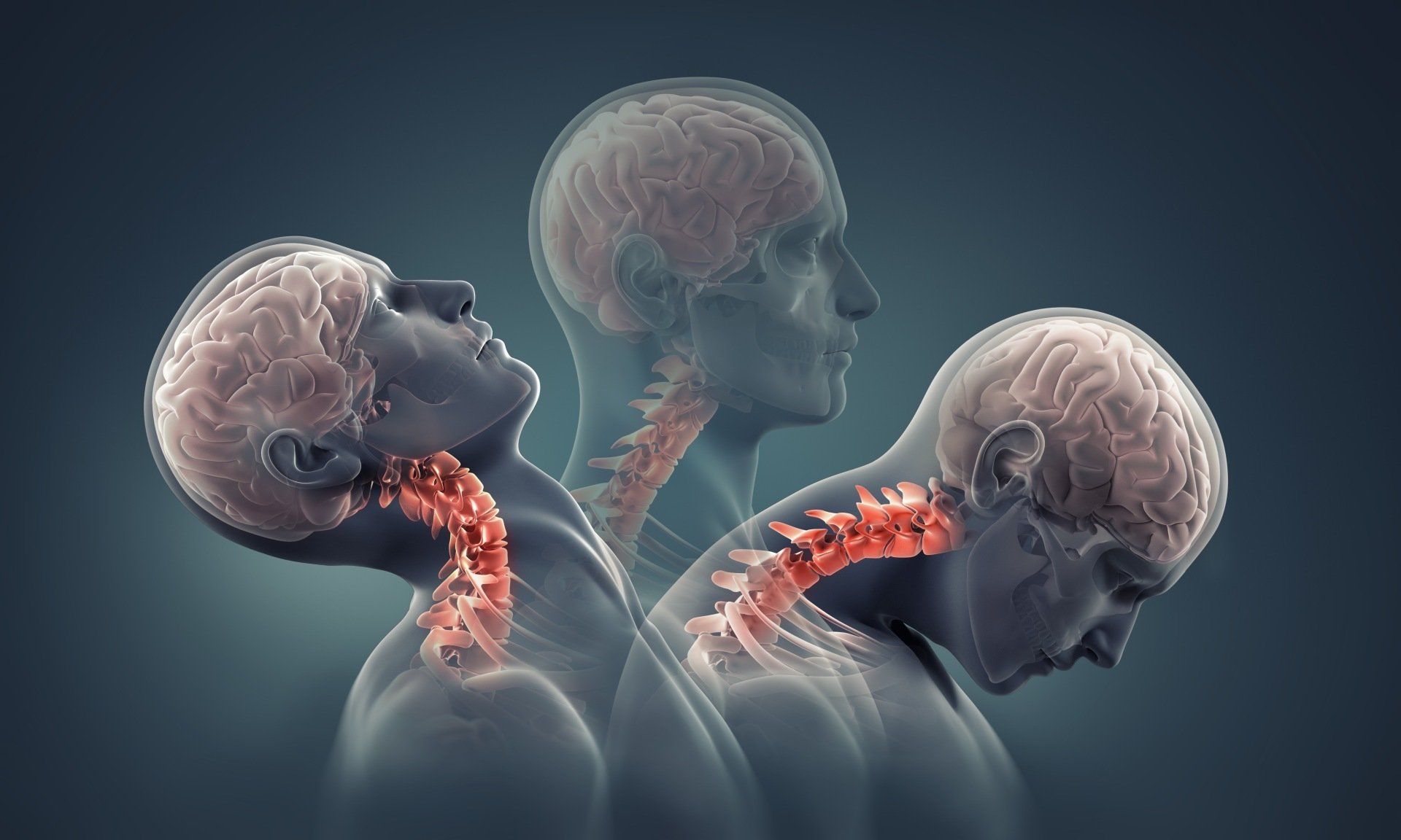
Overview: Whiplash happens when your neck jerks back and forth quickly and violently, causing your spine to bend past its normal range of motion. This can injure the vertebrae of your cervical spine and/or the ligaments and muscles in your neck. Causes: Whiplash is commonly caused by rear-end collision motor vehicle accidents or by contact sports such as football. Symptoms: You may feel pain and stiffness in your neck, shoulders, back, and arms at the time of your injury, or it may begin days later. Treatment Options: Treatment options may include rest, physical therapy, medications and injections.
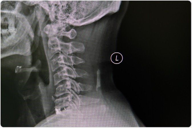
This is a disease of the spine indicating degenerative changes, sometimes referred to as arthritis of the spine. It can happen at any level (cervical, thoracic or lumbar) and results in different types of pain including mechanical and radicular pain. CAUSES Primarily this is caused by aging and normal wear and tear. With time, the components of the spine, including the vertebrae, the discs between the vertebrae, and the joints of the spine rub against one another, causing pain. SYMPTOMS Cervical Spine: neck, shoulder and arm pain. Radicular symptoms may occur including numbness, weakness in the arms or legs. Thoracic Spine: upper chest and abdomen pain, and may cause numbness, tingling and weakness in the legs Lumbar Spine: lower back, buttocks, and leg pain; and may cause numbness, tingling and weakness in the lower leg. This is often times worsened with sitting, twisting, lifting and bending. TREATMENT This varies based on severity of degeneration, and symptoms patients describe. It includes anti- inflammatory medications, physical therapy, water therapy, back braces, spinal injections and in severe cases, surgery.
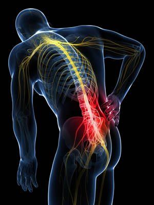
Piriformis syndrome occurs when the piriformis muscle, which is located deep in the buttock, causes pain. The piriformis muscle starts at the lower spine and attaches to the upper thigh bone. It functions to rotate the hip outward. What causes Piriformis Syndrome? Swelling, inflammation and spasm can occur in the piriformis muscle due to trauma, prolonged sitting or repetitive injury such as from sports. The pain may run down the back of the thigh. How is Piriformis Sydrome diagnosed? There is no direct, definitive test for Piriformis Syndrome. It is diagnosed by history and physical exam. Imaging may be ordered to rule out other pathologies. How is Piriformis Syndrome treated? Avoiding the activities that trigger the pain is important. Rest, ice, and heat can be helpful. Physical therapy, stretching, and exercises can also help relieve pain. Anti-inflammatories and muscle relaxers can ease pain. Injections with a steroid or botox can reduce the inflammation, pain, and spasm.
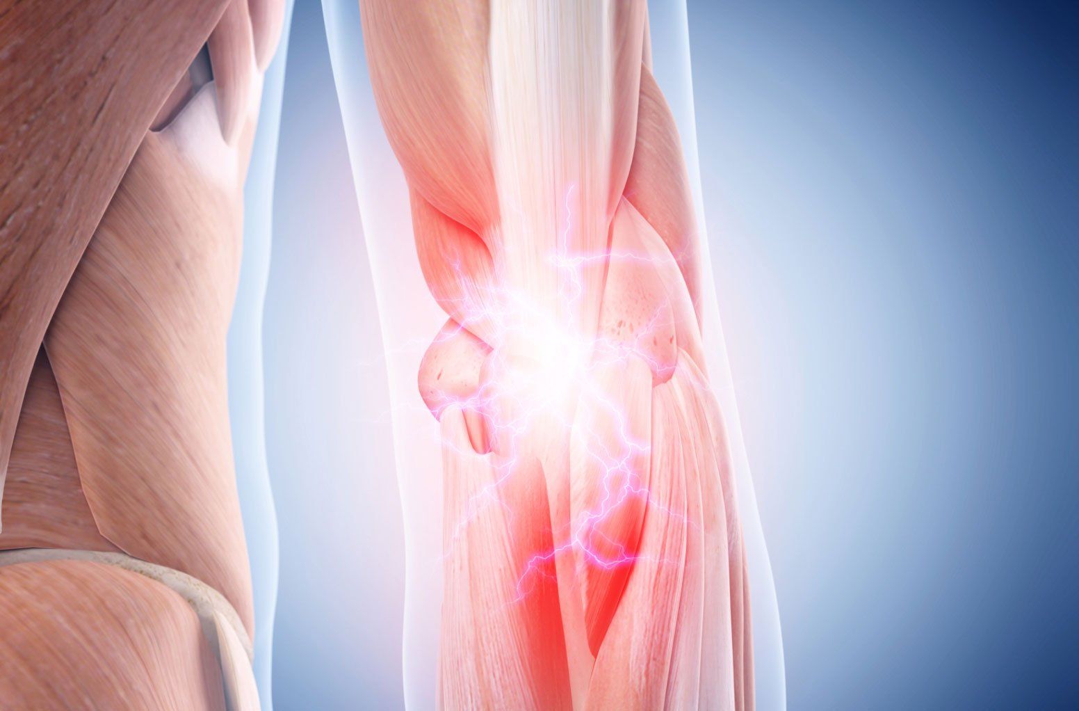
What is medial epicondylitis? Medial Epicondylitis also know as Golfer’s elbow is the inflammation of the tendons that connect the muscles of the muscles of the forearm to the elbow. The pain is felt at the medial epicondyle (the bony bump on the inner side of the elbow). Causes of medial epicondylitis It is caused by repetitive motions, overuse of the arm, and stress on the fore arms which leads to small tears at the flexor tendons triggering pain, weakness, stiffness. Risk Factors: This condition is common among athletes such as golfers and people who play racquet sports. It also is common in persons involved in professions requiring repetitive motions such as carpenters, painters, and computer users. Symptoms of medial epicondylitis There is pain, tenderness, and stiffness at the medial epicondyle that can radiate to the fore arm and wrist. Numbness and tingling in the fingers Weakness of the hand and wrists Difficulty moving the left elbow Treatment Options Rest and avoid activities that require repetitive movement until the pain disappears. Acupuncture therapy, massage therapy and use of an electronic pulse massager can relieve the pain and improve circulation. Ice and a cold compress can help to reduce swelling and inflammation. Physical therapy/Occupational therapy: stretching helps stretch and strengthen the tendons. OTC pain killers and anti- inflammatories can help reduce the pain and inflammation. Topical ointments such as the lidopro ointment to reduced localized pain and inflammation. Steroid injections and PRP injections helps to reduce swelling, inflammation, and can initiate or expedite the healing process. Wrist splint/Brace: can provide additional support and pain relief to reduce muscle strain. Laser therapy that we offer in clinic.
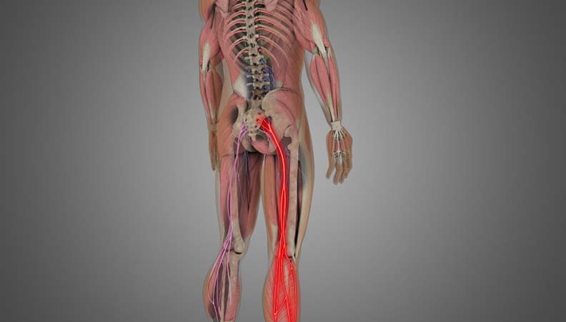
Overview This condition is an irritation or compression of one or more nerve roots in the lumbar spine. Because these nerves travel to the hips, buttocks, legs and feet, an injury in the lumbar spine can cause symptoms in these areas. Causes: Herniated disc: a rupture in the fibrous outer wall of a vertebral disc, which allows the soft nucleus of the disc to bulge outward. This bulge can press harmfully against a nerve root. Degenerative disc disease: when a spinal disc weakens, allowing vertebral bones above and below the disc to shift out of position. The bones can touch, pinching nearby nerve roots. Spinal Stenosis: when bones, discs or joints of the spine degenerate and push into the spinal canal or foramen space. Symptoms Symptoms may include pain, weakness, numbness and tingling, and may vary depending on the level of the injury.
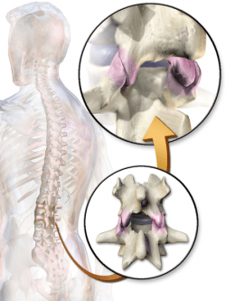
The facet joints are the lateral joints in the spine that connect the vertebrates together. They are lined with cartilage and help promote healthy movement of the spine. They can be damaged through normal aging or through a traumatic injury like whiplash, causing pain or facet joint syndrome. How is facet joint syndrome diagnosed? A practitioner knowledgeable in spinal disorders will take a history, do a physical exam and review imaging. The classic finding of facet syndrome is pain and tenderness over the facet joints with extension (bending backwards). How is facet joint syndrome treated? A conservative nonoperative approach is utilized. This can include physical therapy, anti-inflammatory medications, and low back exercises. In addition, minimally invasive therapies such as injections for diagnosis and treatment may be used. Intraarticular injections involving steroids help reduce pain and inflammation. Medial branch blocks use anesthetic agents on the nerves that arise from the facet joints. If the pain is relieved by these blocks, it is a good sign that a radiofrequency ablation may provide longer lasting relief. This is accomplished by heating the nerves of the facet joints. This can provide pain relief from six months to two years.
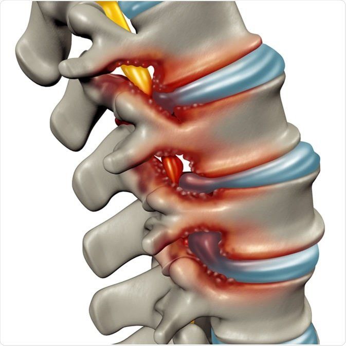
Spinal stenosis is a narrowing of the spaces within your spine, which can put pressure on the nerves that travel through the spine. This occurs most commonly in the neck and low back. Some people may not experience any symptoms, where as others may have pain, numbness, tingling and muscle weakness. Two common causes of spinal stenosis are osteoarthritis, and herniated or bulging discs. Other causes can be from tumors or spinal cord injuries. Spinal stenosis is often diagnosed through MRI and CT scan. In office nerve testing may help determine the health of the nerves affected by the stenosis. Treatment options may include medication, physical therapy and injection therapy, such as epidural steroid injection. If the stenosis is severe enough, or other treatment options fail, surgery may be indicated.
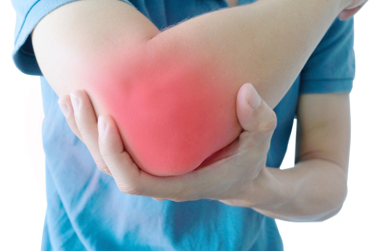
What is it? A nerve problem which is also called Golfer's Elbow or Ulnar Nerve Entrapment. The nerve may become compressed, irritated or stretched on the inside aspect of the elbow causing pain, tingling, numbness and weakness down the inside of the forearm to the 4th and 5th digits. How does it occur? The ulnar nerve passes through the narrow cubital tunnel at the elbow, which is normally cushioned and protected by soft tissue. The area may become damaged by a compression injury. It can become compressed by resting your arm on your armrest for a prolonged time. It can be compressed by a lesion or build up of inflammation. The nerve can become irritated by repetitive use of the elbow such as a golf swing. The nerve can also be damaged by stretching the nerve by bending the elbow for too long such as when you are sleeping, computer work or holding a phone to your ear. How is it diagnosed? Ulnar neuropathy may be diagnosed by symptoms however, confirmation of the diagnosis can be made by electrodiagnostic studies. X-rays may reveal calcium deposits on adjacent bones which can cause irritation to the nerve. MRI of the elbow may be further diagnostic. How is it treated? Icing the inside of the elbow will help decrease inflammation and give relief. Topical and oral antiinflammatories may also help reduce inflammation. Physical therapy is needed when these modalities are not effective. The therapist may use laser light therapy, ultrasound therapy and myofascial release therapy. The therapist will also give you therapy exercises to continue with at home. Wearing a brace over the area at night may be helpful to keep the arm straight while sleeping. Injections and Surgery? When the conservative modalities are not effective, injections with a steroid or biologics may be considered. PRP, or platelet rich plasma injections, can often be helpful. Amniotic fluid injections can be considered to help with inflammation and pain. Surgery can be done in the form of a cubital tunnel release to release the pressure around the nerve by cutting the tissue around the nerve. Ulnar nerve transposition surgery may also be considered to move the ulnar nerve to a place where it is no longer compressed.
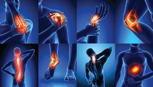
Overview Our goal in Pain Management is to enhance patients' quality of life through decreasing pain, increasing their function, and improvement in the ability to enjoy activities of daily living. We accomplish this by prescribing medication, interventional therapies (epidurals, facet blocks, radiofrequency ablation of nerves), advance therapies (implantation of spinal cord stimulators and/or intrathecal drug delivery systems), have patients exercise (supervised PT, aqua therapy, or home exercises), improve sleep, medications, meditation-sleep hints/rules, referrals to Pain Psychologist/Psychiatrists for cognitive behavior therapy, Functional Restoration Programs, and/or referral to spine/orthopedic specialist and surgeons if needed. Who Can Benefit? Pain management may help you if you have pain that lasts for a long time and/or greater than three months, also known as “chronic pain.” Our goal is to assist/help in reducing spine pain and/or other orthopedic problems, cancer, and/or other long-term illnesses/conditions, as well as injuries, such as industrial/work injury, motor vehicle accidents and post-operative pain. Who Practices Pain Management? Pain management is an intricate network of doctors that have backgrounds in anesthesiology, physical medicine and rehabilitation, neurology, and/or psychiatry. The specialist works closely with your entire healthcare team. Types of Treatment Different types of pain respond to different treatment plans/techniques to include conservative therapies (i.e. chiropractic, acupuncture, massage, physical therapy). Your specialist may recommend injections or the initiation/continuation of conservative treatment therapies, to include lifestyle changes, exercise, and psychological treatments. Conclusion Although some chronic pain conditions may never be cured completely, appropriate pain management/treatment can help reduce your pain and allow you the suitable tools in coping with your pain, leading to an overall improvement in quality of life, function, and tolerance for daily activities.
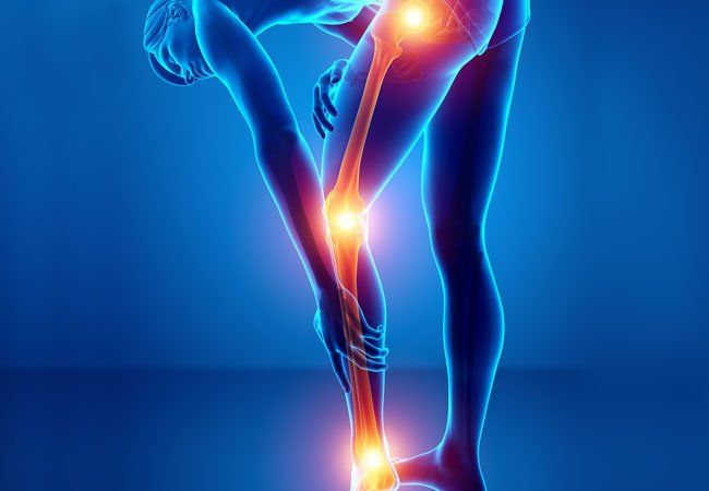
Overview Complex Regional Pain Syndrome (CPRS) is a type of chronic, long-lasting pain affecting one or several limbs or a part of a limb. CRPS has specific attributes of ongoing hypersensitivity, swelling, color and temperature changes in the affected extremity, that was previously injured by trauma or surgery. CRPS is believed to be one of the most underdiagnosed causes of disability, pain, and suffering. With CRPS, you may have unexplained pain that will not go away. It may be severe, and it may even spread. Causes The exact cause of CRPS is not completely understood and/or known. Its origin remains as an abnormal response that your body has to being hurt, almost as a type of overreaction, or like an allergy. It can develop after any kind of trauma or injury to the body (i.e. limb fracture, sprain/strain, blunt trauma, stroke, heart attack, after casting, etc.). Symptoms If you have CRPS, you may feel burning pain, pins and needles, and hypersensitivity or intolerance to light touch. You may also experience increased warmth or coldness, sweating, skin color or nail growth changes or restrictions in use and movement of limb. For example, if you hurt your hand, it may spread to the entire arm or even to the other arm. Your skin may change colors, and may feel warm or cool as well as be overly sensitive to light touch. Treatment It is very important to start treatment ASAP for better results, as CRPS can progress rapidly. Some of the treatment options include: physical therapy, acupuncture, biofeedback, medications, psychotherapy, managing expectations, sympathetic nerve blocks as a diagnostic and therapeutic procedure, neuro and peripheral stimulation, spinal cord stimulation, transcranial magnetic stimulation, and stimulation of the dorsal root ganglion in refractory cases.
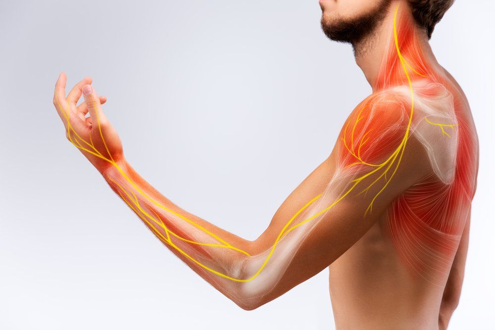
Cervical radiculopathy occurs when one or more of the nerve roots in the cervical spine are being compressed or irritated. This is commonly referred to as “pinched nerve.” This condition can cause neck pain and/or pain radiating down the arms. This can also result in neurological changes including numbness, weakness in the shoulder, arm, hand or fingers. Some of the causes for cervical radiculopathy include a herniated disc, degenerative disc disease and spinal stenosis. Some of the treatments for cervical radiculopathy are physical therapy, medications, steroid injections and surgical interventions.
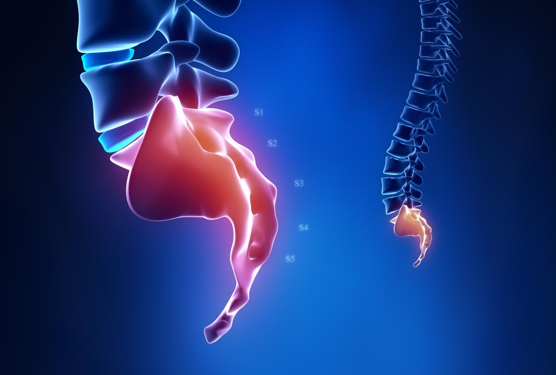
Coccydynia, also known as tailbone pain can arise either from direct trauma or fall, prolonged sitting, or can also occur spontaneously. This pain is caused by inflammation of the tip of the tailbone. The pain symptoms include sensitivity or aching in the tailbone can be made worse with sitting, bowel movements, sexual intercourse. Treatments for coccydynia include using a padded seat cushion, medications, nerve blocks with fluoroscopy guidance, sacral nerve stimulation or surgery to shorten the coccyx.
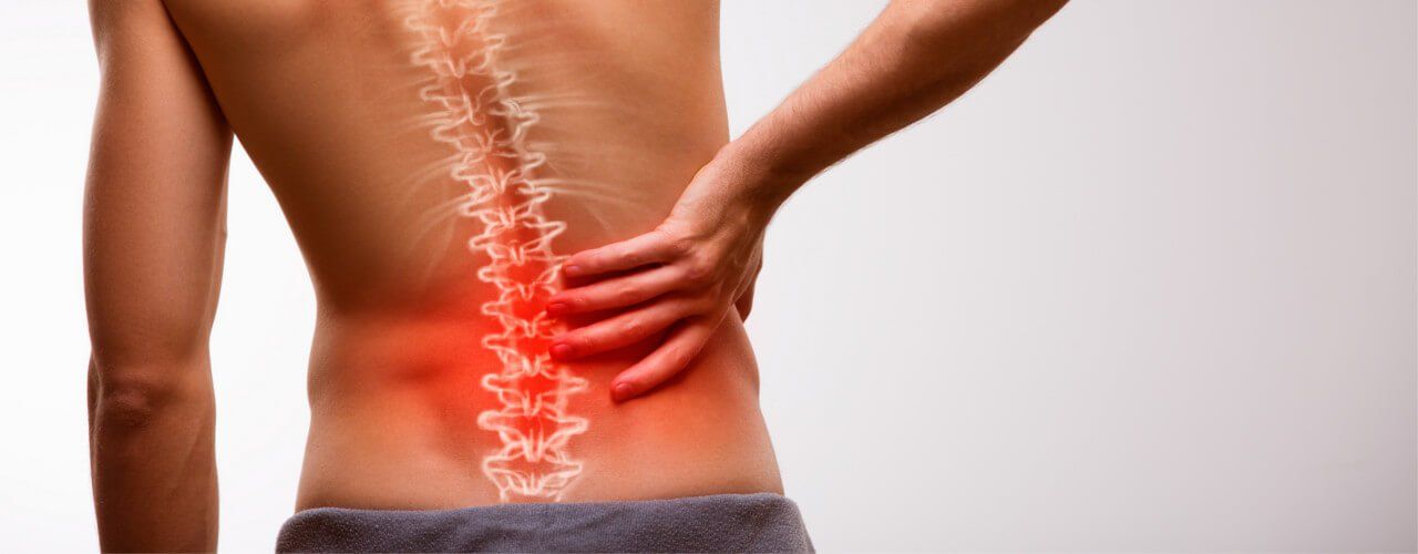
This condition occurs at the disc between the vertebrae of the spine. The discs are soft and let your spine twist and bend and absorb shocks. When damaged, the disc’s soft center pushes through the disc wall and creates a herniated disc. This bulge presses against nerves in your spine. A herniated disc can be caused by wear and tear of aging or from traumatic injury or lifting something heavy. This can cause pain, numbness, tingling or weakness that can be felt in the buttocks, leg or foot. Treatments are based on the injury and include rest, medications, physical therapy, spinal injection with fluoroscopy or surgery.
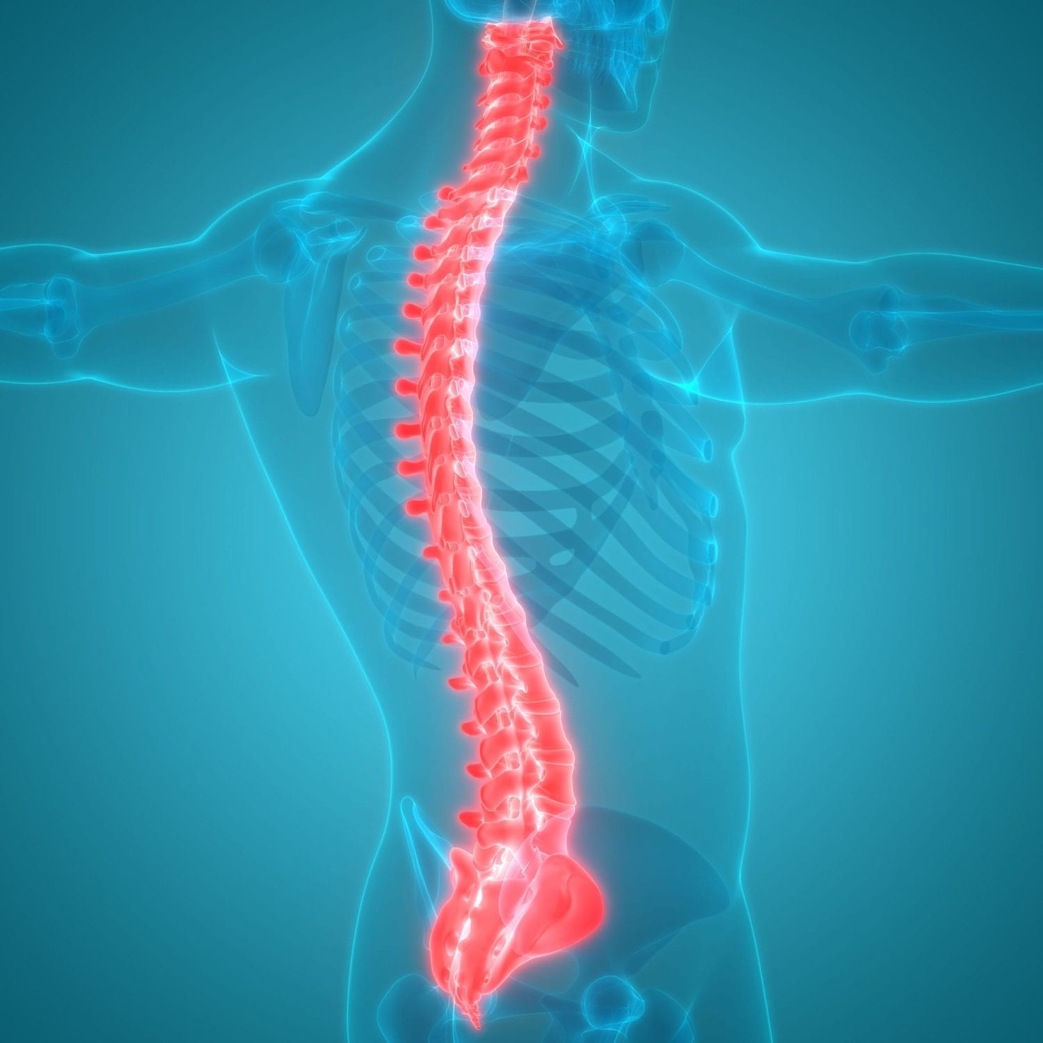
The spinal column is the main support structure of the entire body. It consists of 33 bones, called vertebrae. The spinal column has 5 regions: Cervical spine consists of 7 vertebrae labeled C1 to C7. The first cervical vertebra is called the atlas. The second is called the axis. Together, the atlas and axis form the joint that connects the spine to the skull and allows the head to swivel and nod. Thoracic spine located in the mid back, consists of 12 vertebrae labeled T1 to T12. These vertebrae serve as attachment points for the ribcage. Lumbar spine commonly called the lower back, consists of 5 vertebrae labeled L1 to L5. This is the main weight-bearing section of the spinal column. Sacral region consists of 5 vertebrae that fused together and form a solid bone called sacrum, and they labeled S1to S5 Coccygeal region commonly called the tailbone, consists of 4 small vertebrae. These tiny bones may be fused or separate. Together they form the coccyx, an attachment point for various muscles, tendons and ligaments. The coccyx also helps support the body when a person is sitting. Vertebrae Altogether, the vertebrae of the spine’s five regions support the weight of the body and protect the spinal cord and nerve roots. Each individual vertebra has a complex set of structures necessary to the overall function of the spine. Vertebral Body The main structure of a vertebra is the vertebral body -- a cylinder-shaped section of bone at the front of the vertebra. It is the main weight-bearing section of the vertebra. Vertebral Canal Behind the vertebral body is the vertebral canal. The spinal cord travels through this channel. Spinal Cord The spinal cord is the main bundle of nerve fibers connecting the brain to the rest of the body. The spinal cord ends near the L1 and L2 vertebrae, where it divides into bundles of nerve roots called the cauda equina. The spinal cord typically ends at L2. While many patients worry about spinal cord injury with low lumbar procedures such as epidural steroid injections, there is little to no chance of a direct spinal cord trauma in the low lumbar spine (L4-S1). Nerve Roots Exiting the sides of the spine are nerve roots, thick nerve branches that transmit signals between the spinal cord and the other parts of the body. Pedicles On either side of the vertebral canal are pedicle bones, which connect the vertebral body to the lamina. Lamina The lamina creates the outer wall of the vertebral canal, covering and protecting the spinal cord. Spinous Process Protruding from the back of the lamina is the spinous process. It provides an attachment point for muscles and ligaments that move and stabilize the vertebrae. These are the bumpy ridges that we call feel on our spines when we feel our back. Transverse Processes Transverse processes protrude from the sides of each vertebra. Muscles and ligaments that move and stabilize the vertebrae attach to the transverse processes. Articular Facet The articular facets form the joints where each vertebra connects with the vertebrae above and below it. Each vertebra has four facets (two superior facets and two inferior facets). The facet joints have a covering of cartilage, which allows movement. The two most common causes of chronic pain in the spine are injuries to the facet joints and the intervertebral disc. Intervertebral Disc Between the vertebral bodies are the tough, elastic spinal discs. They provide a flexible cushion, allowing the vertebrae to bend and twist. Each disc has a tough outer wall called the annulus fibrosus and a soft interior called the nucleus pulposus. The two most common causes of chronic pain in the spine are injuries to the facet joints and the intervertebral disc.

Fibromyalgia is a condition which in the past has been a diagnosis of exclusion. This meant that a person was only diagnosed with the condition after all other possible causes of pain were ruled out. In the 1990’s, the American College of Rheumatology (ACR) made the diagnosis of fibromyalgia more clear cut. The ACR’s definition involved assessing an individual who has widespread chronic pain for tender points. 18 specific tender points in the body were described by the ACR. If an individual had 13 out of 18 tender points, then they would meet the criteria for fibromyalgia. Recently the diagnosis of fibromyalgia has been streamlined. The tender point identification is not essential and fibromyalgia is no longer a diagnosis of exclusion. The modern definition requires that a patient have chronic widespread pain that is present for more than 3-6 months. The pain should be symmetric. It should be on the right side of the body, as well as the left side of the body. The pain should be present above the waist and below the waist. It should be present along the spine and in the limbs. The pain of fibromyalgia will typically be associated with muscle stiffness, sleep disturbance and may be associated with irritable bowel syndrome. The above definition of fibromyagia is the most practical way to make an accurate diagnosis. There are other ways to confirm the diagnosis. Brain scans called functional magnetic resonance imaging studies (fMRIs) have shown increased activity in pain centers in the brain in patients with fibromyalgia. Researchers have also shown increased levels of pain neurotransmitters in the cerebral spinal fluid of patient with fibromyalgia. If you have symptoms of chronic widespread pain with morning stiffness and sleep disorder that has lasted for more than 3-6 months, you should discuss your symptoms with your health care providers. There are now FDA approved medication therapy to treat and manage fibromyalgia.
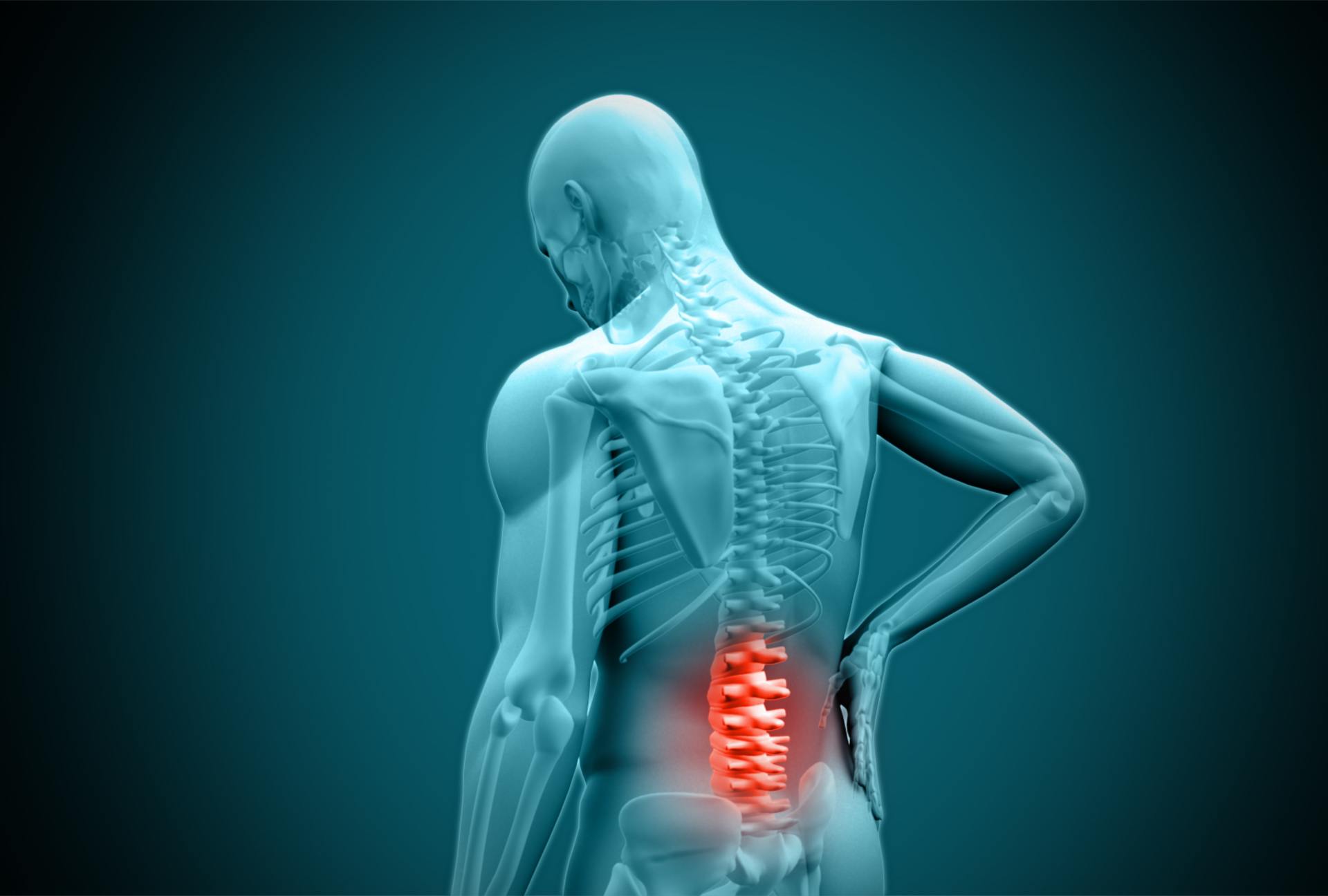
Post-Laminectomy Syndrome, also known as “failed back surgery syndrome,” is a type of chronic pain that can develop in some people after they have had spine surgery. Causes: This pain most often develops after a laminectomy procedure, which is the removal of bone at the rear of your vertebrae. The procedure is done to relieve pressure on your spinal nerves. But after a laminectomy, bone or soft tissue may still press on these nerves, scar tissue may form and spinal joints may be irritated and inflamed causing pain. Symptoms: The symptoms of post-laminectomy syndrome are variable, but often include low back or neck pain, or pain at the site of the surgery. The pain may radiate down to your buttocks or legs and may feel sharp or dull and achy. Some patients can also experience neurologic symptoms which cause pain radiating down the arms. Treatment: If you are having the above symptoms, you should be evaluated by one of the excellent providers at Spine & Nerve Diagnostic Center who are very familiar with treating this condition. Your provider may order an x-ray or MRI of the surgical area. Treatment depends on the cause and the severity of your pain, but may include physical therapy, core stabilizing exercises, stretching, injections or medications. You may also benefit from electrical nerve stimulation, a brace or other treatments.

Degenerative disc disease is a very common cause of low back and neck pain. This pain is caused by wear and tear on the spinal disc. More often than not, degenerative disc disease is associated with chronic low level pain with intermittent episodes of severe pain. The severe episodes can results in weakness, numbness, and radicular pain. This condition can typically be managed with non-surgical treatment methods such as physical therapy, medication management and spinal injections.